
Discovery to Delivery
› Solutions ›Radiotracers or Radiopharmaceuticals
Radioactive tracers are those substances composed of tightly bonded molecules with a radioactive atom. Such substances or tracers often use molecules that normally interact with a specific protein or sugar in the body so medical practitioners can easily determine the exact source of the disease. Others even employ the use of the patient’s own cells which can be seen in cases of a Single Photon Emission Computed Tomography (SPECT) scan wherein healthcare workers radiolabel or add radioactive atoms to a sample of the patient’s red blood cells and then follow its path until they trace the place where it accumulates highly in the body; these areas are called ‘hot spots’.
Radiopharmaceuticals are the approved tracers meeting the standards set the by Food and Drug Administration (FDA). Nuclear medicine physicians are those who specialize the use of such substances to assess body functions, diagnose, and even treat specific diseases. Moreover, the radiopharmaceutical of choice is the thing that determines if the patient must undergo either a SPECT or Positron Emission Tomography (PET) scan.
Single Photon Emission Computed Tomography (SPECT) scan utilizes equipment that provides three-dimensional (tomographic) images showing the distribution of the radiopharmceutical or tracer molecules injected in the patient. These photos are generated by a computer from various amounts of projected images recorded during the scan with different body angles. The computer uses a set of cameras mounted on a rotating gantry which in turn, moves the detectors in a tight circle around the pallet where the patient lies on a supine position, motionless.
The radiopharmaceuticals used in SPECT scan must emit gamma rays injected to the patient. These rays move at a different wavelength than visible light. Also, the detectors used during SPECT scan can easily detect these gamma rays.
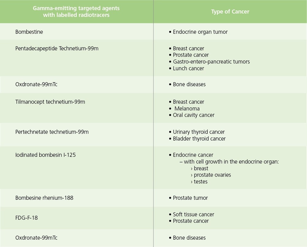
There have been a large number of compounds radiolabelled with gamma-emitting substances to allow imaging of different types of cancer.

Table 1. Gamma-emitting radiopharmaceuticals for diagnostic imaging different types of cancer and diseases.
SPECT scans are primarily used to diagnose and track the progression of diseases such as heart disease, bone disorders, gall bladder and even intestinal bleeding. Recent studies suggest that SPECT agents can also aid in diagnosing Parkinson’s disease in the brain.
Positron Emission Tomography (PET) scan is another type of powerful imaging technique utilizing radiopharmaceuticals to generate three-dimensional images. It uses quantitative information based on the distribution of positron emitters (PET radiopharmaceuticals) injected or administered reflected by the patient’s body.
PET studies produce images representing the distribution or the pattern of distribution as taken up by the body which can vary based on its current physiologic, pharmacologic, and biochemical state. As such, PET scans are able to provide information of the bodily functions of a patient.
The difference between the SPECT and PET scan lies on the type of radiopharmaceuticals or radiotracers, used. While SPECT utilizes substances emitting gamma rays, PET uses radiotracers that produces small particles called positrons during their decay.
Positrons have the ability to react with the body’s electrons as they are oppositely charged (positron is positively charged while electrons are negative). When these particles combine, they annihilate each other, an act that produces a small amount of energy in the form of photons, and these shoot off in different directions. The PET scanner then measures these photons and utilizes them to create images of the internal organs of the patient.
Glucose utilization is dependent on the intensity of cellular and tissue activity and is greatly increased in rapidly dividing cancer cells. One fact is that, the degree of a cancer’s aggressiveness can be measured as it is paralleled by their rate of glucose utilization. For the past decade or so, glucose molecule called F-18 labeled deoxyglucose or FDG, has been shown to be the best tracer to detect cancer and its metastatic spread in a patient.
The main purpose of PET scan is to detect and monitor and detect metastases as well as track the progression of cancer in response to treatment.
A combination instrument that produces both PET and CT scans of body regions one exam has become the primary tool for cancer staging worldwide.
Moreover, recently a PET probe was approved by the USFDA for the accurate diagnosis of Alzheimer’s disease.
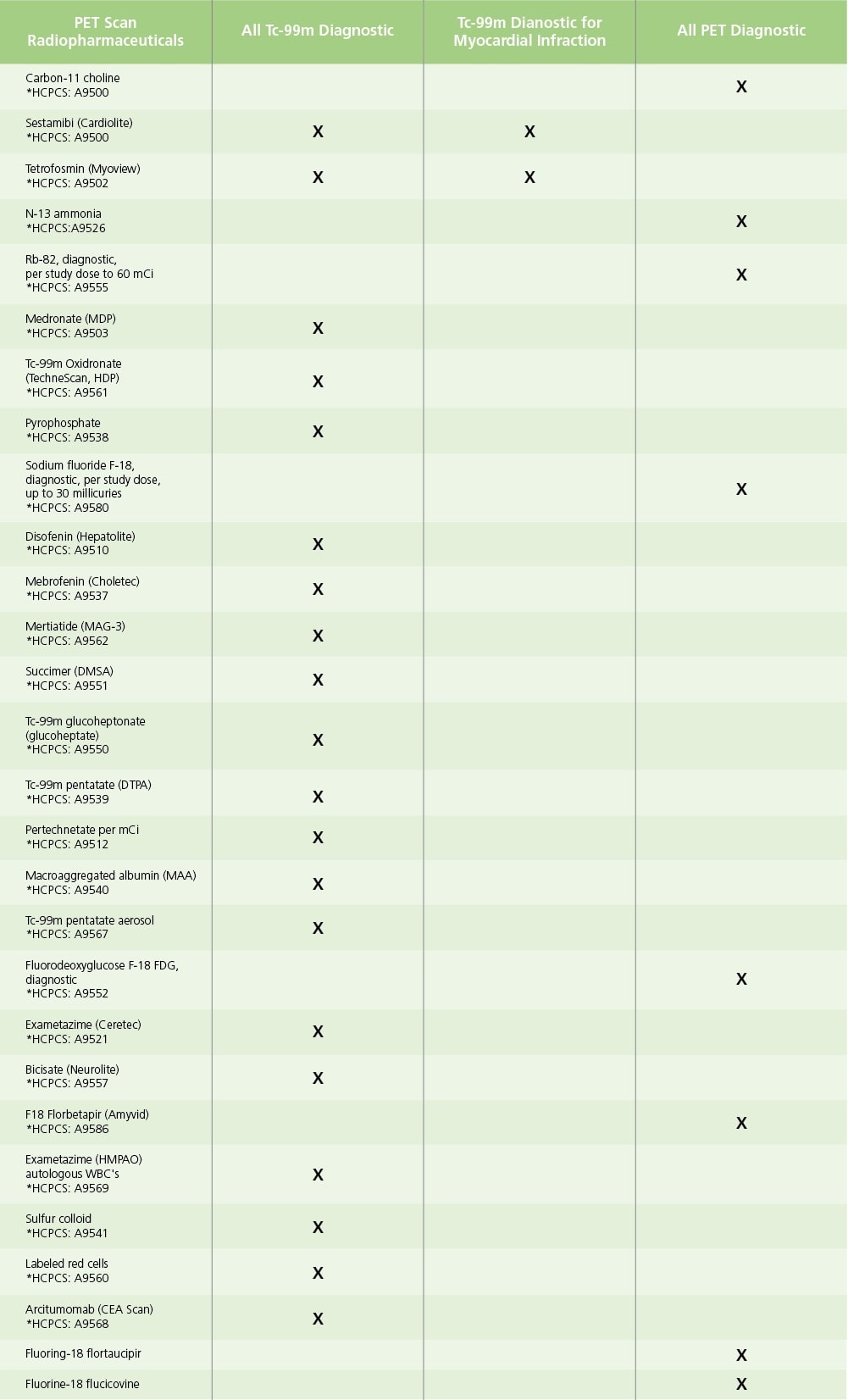
 *Healthcare Common Procedure Coding
Table 1. Sample of Approved Radiopharmaceuticals for PET Scan.
SPECT scans are primarily used to diagnose and track the progression of diseases such as heart disease, bone disorders, gall bladder and even intestinal bleeding. Recent studies suggest that SPECT agents can also aid in diagnosing Parkinson’s disease in the brain.
References:
Recommended Products
Sign up to our newsletter and receive the latest news and updates about our products!
